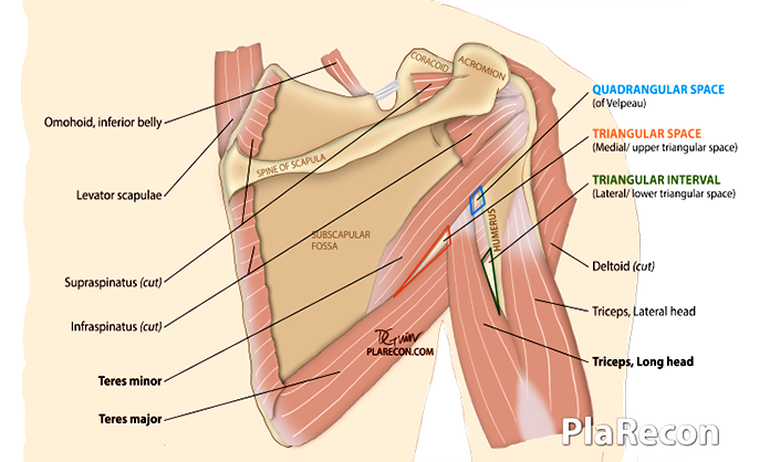
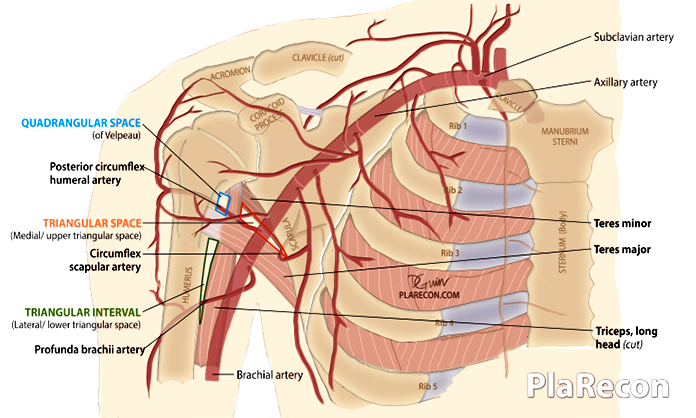
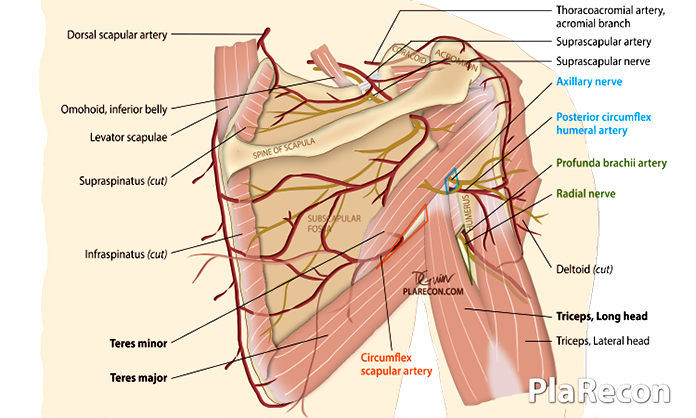
Three intermuscular spaces are present in the scapular region/ arm:
- Triangular Space – TS (aka– Medial or Upper Triangular space, Foramen omotricipitale),
- Quadrangular Space – QS (aka– Quadrilateral space of Velpeau, Foramen humerotricipitale),
- Triangular Interval – TI (aka– Lateral or Lower Triangular space, Triceps hiatus).
[Color-code used in this post and the illustrations: TS–red, QS–blue, TI–green.]
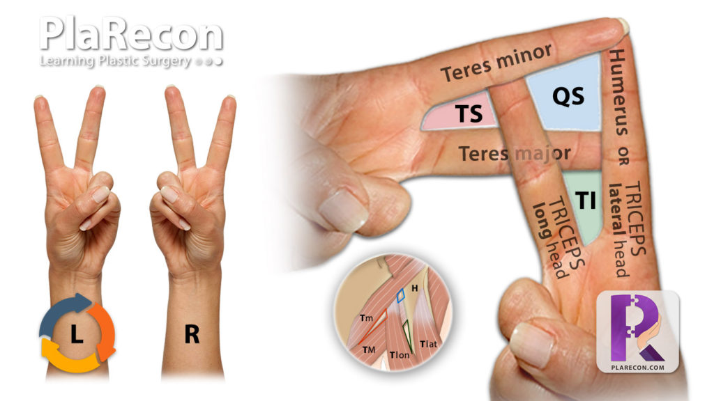
💡 Mnemonic for Boundaries/Borders
- 💡 Double ‘Victory’ sign! [Details in the image below]
- Among the Teres muscles- Major is heavier, hence sunken lower down (T. major is inferiorly placed),
- Among the Triceps heads- Long head is represented by the Longest (i.e. middle) finger, Voila!

The structures forming the boundaries or borders and contents of these spaces are described below.
Triangular Space
Boundaries/Borders
- Superior: lower margin of Teres minor,
- Inferior: upper margin of Teres major,
- Lateral: medial margin of the Long head of Triceps.
Content (1 artery)
- Circumflex scapular artery (is a posterior branch of Subscapular artery, which is a branch of the third part of the Axillary artery)
Quadrangular Space
Boundaries/Borders
- Superior: Inferior margin of Teres minor,
- Inferior: Superior margin of Teres major,
- Medial: Medial margin of the Long head of Triceps,
- Lateral: Medial margin of shaft of Humerus.
Contents (1 Artery, 1 Nerve)
- A- Posterior circumflex humeral artery (its the last branch of the Axillary artery)
- N- Axillary nerve (which after passing through this quadrangular space divides into anterior and posterior branches to innervate the Teres minor and Deltoid and provide sensation to the upper lateral arm- Regiment badge area).
Triangular Interval
Boundaries/Borders
- Superior: Inferior margin of Teres major,
- Medial: Lateral margin of the Long head of Triceps,
- Lateral: Medial margin of shaft of Humerus or the Lateral head of Triceps.
Contents (1 Artery, 1 Nerve)
- A- Profunda brachii artery
- N- Radial nerve (which passes through the Triangular Interval with its first branch- to the long head of Triceps- usually given off 1 cm proximal to the inferior margin of Teres Major).
Clinical applications
Some clinical conditions (with relevance to Plastic Surgery) where in-depth knowledge of the anatomy of these spaces is required:
Brachial plexus reconstruction
Distal nerve transfer for restoration of shoulder function in C5,6 Upper Brachial Plexus Injury (BPI): Somsac transfer- The nerve to the long head of Triceps (first branch of the radial nerve- in or below Triangular interval) to the anterior division of the Axillary nerve in the Quadrangular space.
Tendon transfers for shoulder function in Birth brachial plexus palsy (BBPP) using the conjoint tendon (i.e. Lattisimus Dorsi+Teres Major: that forms the posterior axillary fold).
Scapular and Parascapular flap
These flaps are based on transverse and descending cutaneous branches of the Circumflex scapular artery after passing through the Triangular space.
Quadrangular space syndrome (QSS)
Aka Quadrilateral space syndrome is caused by compression of the axillary nerve and/or the posterior circumflex humeral artery in the Quadrangular space by fibrous band(s) with or without surrounding muscle hypertrophy or rarely dilated veins.
Triangular Interval Syndrome (TIS)
It is a compressive neuropathy of the Radial nerve in the Triangular Interval leading to upper limb radicular pain.
If you found this post useful subscribe below for more such articles & subscribe to our YouTube channel for video tutorials. You can also find us on Facebook, Twitter and Instagram.
References:
- Netter’s Atlas of Human Anatomy, 6Ed (2014)

Tutorials & tips in Plastic Surgery
+ Weekly updates of high-quality webinars!


Thanks doc, medical school loves you
This is so helpful! Thank you!